|
Prokaryotic Cells
unicellular organisms
Eukaryotic Cells
multicellular organisms
unicellular and multicellular organisms
Viruses
nonliving
made of nucleic acids and proteins
Assignment
1. Read Chapter 7
pages 152-166
(Hint: study the pictures)
2. Do the Directed Reading Questions:
7.1 Questions
7.2 Questions
7.3 Questions
7.1 PowerPoint
7.2 PowerPoint
7.3 PowerPoint
Structures and Functions Notes
|
1. Read the microscope magnification worksheet and complete the table in your notebook
2. Use a microscope to see:
- prokaryotic cells: bacteria
- unicellular eukaryotic cells: protists
- multicellular eukaryotic cells: animals
3. Draw and label the cells at different magnifications in your notebook
4. Draw a scale bar and estimate the size of the cell in your notebook
- prokaryotic cells: 40X, 100X, 400X
- protist cells: 40X, 100X, 400X
- animal cells: 40X, 100X, 400X
|
Cell and Virus Models
Use the textbook and the Cells Alive interactive animation
- Draw and label a diagram of each cell in your notebook
- Draw and label the structures of each cell in your notebook - see below
- Draw and label the structures of a virus in your notebook
Draw and label these structures:
prokaryotic cell structures (Chapter 20)
- cell wall
- plasma membrane
- pilus (pili plural)
- flagellum (flagella plural)
- ribosome
- cytoplasm
- nucleoid
- plasmid
virus structures (Chapter 20)
- nucleic acid (DNA or RNA)
- capsid
- *envelope (optional)
- *tail fibers (optional)
|
Cell and Virus Models (cont.)
Draw and label these structures:
eukaryotic animal cell structures
- plasma membrane
- cytoskeleton
- nucleus
- nucleolus
- chromatin
- nuclear membrane
- cytoplasm
- rough endoplasmic reticulum
- smooth endoplasmic reticulum
- Golgi apparatus
- vesicle
- vacuole
- mitochondrion (mitochondria plural)
- lysosome
- peroxisome
- centrosome
- centriole
- ribosome
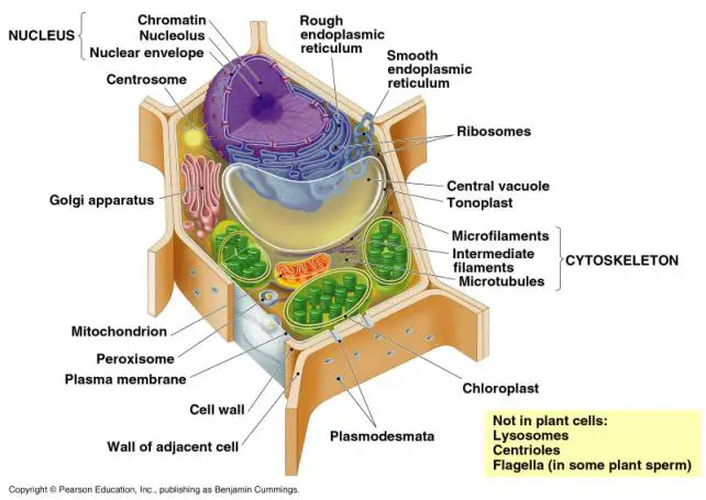
eukaryotic plant cell structures
1-18 above
19. cell wall
20. chloroplast
*12. central vacuole
|
|
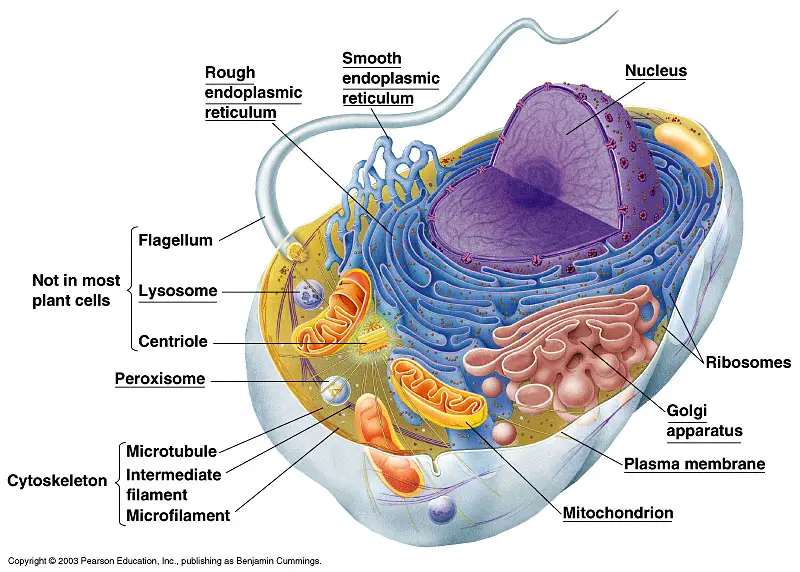
Functions of Cell Structures - Animal Cell
- organelle: any cell structure with a membrane (“little organ”)
1. plasma membrane (cell membrane): controls movement of substances and information into and out of the cell
2. cytoskeleton: keeps the shape of the cell and holds and moves the organelles
3. nucleus: organelle that protects the DNA
4. nucleolus: inside the nucleus, makes ribosomes
5. chromatin: DNA wrapped loosely around proteins in the nucleus
6. nuclear membrane: controls movement of substances into and out of the nucleus
7. cytoplasm: liquid that fills the cell, mostly water with many molecules, compounds, and ions dissolved in it where a lot of chemical reactions happen
8. rough endoplasmic reticulum (RER): folded membrane near the nucleus that has ribosomes attached to it, helps make proteins and send them out of the cell
9. smooth endoplasmic reticulum (SER): folded membrane that makes lipids
10. Golgi apparatus: organizes and packages proteins made by the RER into vesicles to transport out of the cell
11. vesicle: a small membrane ball used to hold and move substances
12. vacuole: a large membrane ball for storing substances
13. mitochondrion (mitochondria plural): produces ATP energy from food molecules
14. lysosome: a vesicle that contains enzymes that recycle old cell structures
15. peroxisome: a vesicle that contains enzymes that break down cell waste
16. centrosome: organizes microtubules for cell division and building cilia and flagella
17. centriole: microtubules inside the centrosome that make new microtubules
18. ribosome: makes proteins
|
Eukaryotic Animal Cell
|
|
Prokaryotic Cell (bacteria)

Structures and Functions
-
cell wall: hard protective outside the plasma membrane
-
plasma membrane: controls transport in and out of the cell
-
pilus (pili plural): used to attach to things
-
flagellum (flagella plural): used for movement (like a tail for swimming)
-
ribosome: makes proteins
-
cytoplasm: liquid that fills the cell, where the chemical reactions happen
-
nucleoid: region where the DNA is (no nucleus)
-
plasmid: small circular piece of DNA
|
Virus (not alive, not a cell)

Structures and Functions
-
nucleic acid (DNA or RNA): information for making more viruses
-
capsid: protective protein coat that holds the nucleic acids, helps attach and enter cells
-
envelope (optional): lipid layer outside the capsid that helps attach to cells
- tail fibers (optional): used to attach to cells and insert the nucleic acid into the cell
|
|
Assessments
|
Notebook - Microscopy
1. Microscope magnification table (4 points)
2. Drawings of cells
- 9 drawings - clear and accurate (4 points)
- magnification and cell labels - for example: "Protist cell 100X" (4 points)
- scale bars - accurate (4 points)
- cell size - accurate (4 points)
4 points = complete, 3 points = mostly complete, 2 points = partly complete, 1 point = very little complete
Total possible = 20 points
|
Notebook - Cell and Virus Models
- four models (4 points)
- structures (4 points)
- labels (4 points)
4 points = complete, 3 points = mostly complete, 2 points = partly complete, 1 point = very little complete
Total possible = 12 points
|
Cell Structure and Function Quiz
(Textbook 7.1, 7.2, 7.3)
|
|
Read Chapter 8
pages 176 - 186
Do the Directed Reading Questions
8.1 Questions
8.2 Questions
8.3 Questions
8.1 PowerPoint
8.2 PowerPoint
8.3 PowerPoint
Homeostasis: keeping stable conditions inside the cell or organism despite a constantly changing environment
Example: membranes control the movement of water and other substances into and out of cells and organelles. Membranes maintain stable concentrations of water and substances inside cells and organelles.
|
Cell Membrane Structure
Phospholipids
The cell membrane is made of a double layer of phospholipids.
- Phospholipids are special lipids that have a polar molecule attached at one end
- The phospholipid has two fatty acids at the other end
- The phospholipid has a polar "head" and a nonpolar "tail"
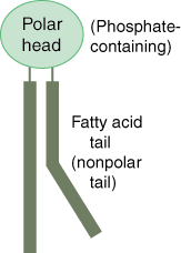
- Outside the cell is mostly water
- Inside the cell is mostly water
- The phospholipids are arranged in two layers so that the polar "heads are facing outside to the water and inside to water
- The nonpolar "tails" are inside the membrane
Proteins
Membrane proteins are part of the cell membrane.
There are four kinds of membrane proteins
- Cell surface markers - identify the cell
- Receptor proteins - receive signals from outside the cell
- Enzymes - help with chemical reactions inside the cell
- Transport proteins - move substances into and out of the cell
|
Cell Membrane Function
The cell membrane controls the movement of substances and information into and out of the cell.
1. Passive Transport
Solution = solute + solvent
The solute is mixed in the solvent to make a solution.
The solute is any substaqnce dissolved in water.
The amount of solute can be the same inside and outside the cell = equilibrium.
When there is no equilibrium, there is a concentration gradient = more solute on one side than the other.
Diffusion = the movement of solute from high concentration to low concentration.
Facilitated diffusion = the movement from high to low concentration through a channel protein or carrier protein. Large and polar compounds cannot move through the phospholipids, so they go through pores made by channel proteins or they are carried across the membrane by carrier proteins.
Video
Osmosis = the movement of water from high to low concentration of water.
Diffusion, facilitated diffusion, and osmosis are passive transport because no energy is used to move the substances.
|
Cell Membrane Function (continued)
2. Active Transport
Active transport requires energy because substances are moving against the concentration gradient from low to high.
pumps: some carrier proteins use ATP energy to pump substances against the concentration gradient.
Video
vesicles: large substances are moved in vesicles.
Endocytosis is bringing large substances into the cell by making a vesicle from the cell membrane.
Exocytosis is moving large substance out of a cell by joining a vesicle to the cell membrane.
Video
3. Communication
Information moves into and out of the cell through the cell membrane.
Signals are messages sent between cells. Signals can be chemical or electrical.
Chemical signals can affect one or many cells.
Electrical signals are specific to one cell or group of cells.
Receptor proteins receive signals and pass the message through the membrane. Receptor proteins have specific shapes, so they only receive one kind of signal. A cell membrane has 100s or 1000s of different receptors.
Response: when a receptor protein gets a signal, it can cause three things to happen to the cell:
- permeability change: passive transport proteins can open or close, and active transport proteins can be turned on or off.
- enzyme activation: an enzyme can begin to do chemical reactions inside the cell.
- second messenger: another chemical signal can change activities in the cytoplasm or the nucleus of the cell.
|


Comments (0)
You don't have permission to comment on this page.